Congenital Viral Infections Slide Set
Rubella
Rubella was first described by two German physicians in the mid-eighteenth century and recognized as a distinct disease in 1881 by the International Congress of Medicine. Its association with congenital defects was not recognized until 1941 when Sir McAlister Gregg reported 78 babies with congenital cataract born after an extensive epidemic of rubella in Australia in 1940. These findings were subsequently confirmed by numerous retrospective and prospective studies.
A. Properties
Non-arthropod-borne togavirus
only member of the genus Rubivirus
ssRNA enveloped virus, 60nm in diameter, nucleocapsid 33nm
symmetry thought to be icosahedral but virus particle has a pleomorphic appearance.
+ve 40S RNA consists of 3 structural proteins; E1 E2 membrane bound glycoproteins, and C capsid protein
Only one serotype of Rubella virus
E1 has 6 distinct antigenic determinants; 4 associated with haemagglutination, and 2 with neutralization
Rubella grows in a wide range of cell lines. It induces a CPE
only in continuous cell lines such as RK13 (rabbit kidney) and
Vero. Immunofluourescense is used to identify the presence of the
virus in culture. Rubella virus infects various primates and
other mammals e.g. monkeys, chimpanzees, rabbits and mice.
Attempts to reproduce the teratogenic effects of Rubella virus in
animal models have produced inconsistent and irreproducible
results.
Rubella has a worldwide distribution. Before the introduction of vaccination outbreaks tend to occur Spring and Summer. Infection is uncommon in preschool children but outbreaks involving school children and young adults were common. In general, about 50% of 10 year olds have rubella antibodies. 80% of women of childbearing age were found to be immune in the prevaccination era. In the UK major epidemics occur every few years. The last one was in 1978. Children 3-10 yrs are most frequently affected. Despite the vaccination program 5-10 % of women reach child bearing are susceptible to Rubella infection.
C. Clinical and Virological Features
Rubella is transmitted by the respiratory route. The
incubation period is 13 to 20 days, during which a viraemia
occurs and virus disseminates throughout the body. In children
the onset is abrupt with the appearance of the rash. In adults a
prodromal phase may be present with fever and malaise for a day
or two before the rash develops. The rash is typically a
maculopapular rash, which first appears on the face and then
spreads to the trunk and the limbs. The rash seldom lasts more
than 3 days. The exact mechanism of how the rash is induced is
uncertain but an immunopathological mechanism may be present.
Lymphadenopathy may precede the rash by up to a week and persists
up to 2 weeks after the rash has gone.
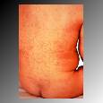
An example of the maculopapular rash found in rubella
Joint involvement is the commonest complication and occurs in up to 60% of adult females. The fingers, wrists knees and ankles are most frequently affected. The arthralgia lasts usually 3-4 days. Arthralgia is rare in males and prepubertal females. An encephalitis develops in 1 / 10000 but the prognosis is good. Trombocytopenic purpura may occur which may present as purpuric rash, epitaxis, haematuria and GI bleeding. In 25% the infection is subclinical It is difficult to diagnose rubella on clinical evidence alone. A similar disease may be caused by parvovirus, arboviruses and enteroviruses. Therefore, a laboratory diagnosis of rubella is essential. Patients are potentially infectious over a prolonged period. Virus can be isolated from nasopharyngeal secretions ( and occasionally from faeces and urine ) 7 days before and up to 7 days after the appearance of the rash. Virus in stools and urine do not play an important role in the transmission of the virus. Viraemia is present for a week before the onset of the rash but as rubella antibodies develop the viraemia ceases.
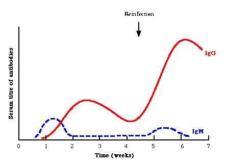
Reinfection - Natural infection is followed by a high level of protection from reinfection. However reinfection can occur which is generally asymptomatic. Reinfection in pregnancy is thought to pose minimal risk to the fetus. Reports of fetal infection are exceedingly rare. In reinfection, the IgG is highly elevated whilst IgM may be demonstrable, giving equivocal results. It may now be possible to distinguish reinfection from primary infection by examining the avidity of specific IgG (IgG from patients with reinfection have a higher avidity)
1. Pathogenesis;- Rubella virus enters the fetus during the maternal viraemic phase through the placenta. The damage to the fetus seems to involve all germ layers and results from rapid death of some cells and persistent viral infection in others. Chromosomal aberrations and reduced cell division are present. The fetus is almost invariably infected if the mother is infected during the first trimester. After the first trimester, the virus is isolated infrequently from the neonates, probably because fetal immune mechanisms can be activated and infection can be terminated. Following intrauterine infection in early pregnancy the virus persists throughout the gestation and can be isolated from most organs at autopsy. Virus can also be recovered from nasopharyngeal secretions, urine, stools and CSF from survivors. However by the age of 3 months the proportion excreting virus has declined to 50-60% and by 1 year, 10%. The mechanism of virus persistence is not known but may be due to defects in cell-mediated immunity.
2. Risk to the fetus
The risk of major malformations following infection in the first trimester varied from 10% to 54%, the risk being greatest in the first 8 weeks of pregnancy (Hanshaw et al 1985). However this data was compiled from prospective studies which were carried out before laboratory diagnosis of rubella infection became available. More recent studies suggest that the actual risk of major fetal damage is much higher than realized. Cardiac and eye defects are more likely to result when maternal infection is acquired during the first 8 weeks of pregnancy i.e. during the critical phase of organogenesis. Whereas retinopathy and hearing defects are more evenly distributed throughout the first 16 to 20 weeks of gestation.
Rubella virus is seldom isolated from infants whose mothers acquired rubella after the first trimester. However rubella- specific IgM can be detected in a high proportion of these infants which means that they were infected. Major abnormalities are very rare because organogenesis is complete by 12 weeks and the immune response may be more developed. Deafness and retinopathy (which does not affect vision), are likely to be the only abnormalities associated with post first trimester rubella. Deafness is usually the sole clinical manifestation of fetal infection occurring between 13 and 16 weeks.
3. Clinical Features
The clinical features of the congenital rubella syndrome (CRS) may be categorized as transient, developmental and permanent.
Transient;- IUGR, thrombocytopenic purpura,
hepatoslenomegaly and haemolytic anaemia. These
abnormalities are present during the first few weeks of
life and are not associated with permanent sequelae.
Transient bone lesions occur in 20% of congenitally
infected infants. 25% have a meningoencephalitis which
may or not leave neurological sequelae. Jaundice is
commonly present.
Developmental;- Sensorineural deafness, mental
retardation, insulin-dependent diabetes. Developmental
defects may take months before they become apparent but
persists permanently. Congenital rubella remains the
commonest cause of congenital deafness in developed
countries. Rubella deafness may be unilateral or
bilateral and varies considerably in severity. IDDM is
actually a common manifestation of CRS ( up to 20%).
However onset may be delayed till adolescence or
adulthood. Autoimmune mechanisms may be involved. Between
3 - 12 months some infants develop a rubbelliform rash,
persistent diarrhoea and pneumonitis which is referred to
as "late onset disease". This carries a high
mortality.
Permanent;- Heart defects (patent ductus, VSD, pulmonary valve stenosis), eye defects (retinopathy, cataract, microopthalmia, glaucoma, severe myopia), CNS defects (microcephaly, psychomotor retardation).
In the early sixties before the advent of vaccination, a large outbreak of CRS occurred in America. A follow-up study was conducted 25 years later and it was found that one-third of those affected were leading normal independent lives, one-third had to live with their parents, and one-third were institutionalized. Late sequelae, especially those affecting the heart were commonly seen.
1. Serological diagnosis of rubella infection - Serology
is the mainstay of diagnosis of rubella infection. A recent
rubella infection can be diagnosed by (1) detection of
rubella-specific IgM, (2) rising titres of antibody in HAI and
ELISA tests, and (3) seroconversion. It is essential to obtain
accurate information relating to the date and time of exposure,
the date of onset of illness. A history of previous rubella
vaccination as well as previous results of rubella screening
tests. Blood should be collected from pregnant women with
features of rubella-like illness as soon as possible after onset
of symptoms. A significant rise in HAI antibodies can often be
demonstrated. However rubella-specific IgM is the test of choice
for demonstrating current infection. It has been shown though
that low and transient levels of IgM can be detected in cases of
reinfection. Furthermore, low levels of rubella IgM may persist
for a few months to 4 years following rubella vaccination.
Typical serological events following acute rubella infection. Note that in reinfection, rubella-specific IgM is usually absent or present at a low level transiently
Haemagglutination inhibition (HAI) assay remains the
mainstay test for diagnosis of rubella infection. HAI Abs
may be detected on the first day of the rash and rise
rapidly to peak titres. It may be difficult to
demonstrate a significant rise in titre unless the serum
is obtained within the first few days of the onset of
illness. Before testing by HAI, sera must be treated to
remove red cell agglutininins and serum lipoproteins. It
is current practice to confirm a serological diagnosis by
HI with testing for rubella IgM.
EIA and RIA have replaced HAI for the diagnosis of
rubella in some laboratories. Occasionally SRH can be
used for diagnostic purposes provided the wells are of
consistent size and a measured volume of serum is added
to each well.
Detection of rubella-specific IgM by EIA or RIA. The most sensitive and reliable techniques in use are tM - antibody capture ELISA and radioimmunoassay). IgM antiglobulins such as Rheumatoid Factor can seldom cause false positive results as can heterophil antibodies. Indirect ELISA and RIA has also been developed but these are not as sensitive as direct ELISA.
2. Serological techniques used for rubella antibody screening;- Single Radial Haemolysis (SRH) and latex agglutination (LA), and .ELISA are used for screening for immunity against rubella. SRH is reckoned to be slightly less sensitive than LA or ELISA. False negative results may occur with SRH due to the interference with red cell lysis by a "blocking factor" which may be removed by absorbing the sera with erythrocytes from the same species used in SRH tests. ELISA is now the test of test in many laboratories but it is considerably more expensive than the SRH. An antibody titre of equal or greater than 15 IU/ml is regarded as being immune to rubella. However, there is some controversy as to the 15 IU/ml cutoff since it was arrived at empirically in the first place. It is quite clear that lower levels of antibody, such as 10 IU/ml would probably be protective as well. HAI is not used for rubella antibody screening because it is not sensitive enough.
3. Virus isolation and identification;- Virus isolation is now seldom used for diagnosing postnatally acquired rubella infection. It may still be useful in diagnosing congenital acquired disease and in determining the duration of virus excretion in these infants since they may transfer infection to susceptible adults. The specimen should minced or mixed with culture medium and the supernatant is then inoculated into culture medium. RK13 cell cultures are used exclusively in the UK for virus isolation. SIRC cells are used in Scandinavian countries. Rubella virus is fastidious and produces CPE in RK13 cells under carefully controlled conditions. The virus is more rapidly identified using IF techniques. Inoculation of specimens into Vero cells followed by passage into RK13 or SIRC cells, in which the virus can be identified by its characteristic CPE or by IF represents the most sensitive method for rubella isolation available.
4. Detection of viral nucleic acid:- RT-PCR is being increasingly used for the diagnosis of rubella infection, especially congenital infection.
4. Diagnosis of congenital acquired infection
The diagnosis of congenitally acquired rubella is made by;
The presence of rubella IgM in cord blood or serum samples taken in infancy.
Detection of rubella antibodies at a time when maternal antibodies should have disappeared (approx.6 months of age)
Isolation of rubella virus or detection of rubella virus nucleic acid from infected infants in the first few months of life.
The detection of rubella-specific IgM in cord blood or infant sera is the method of choice for the diagnosis of congenital rubella. Specific IgM has been demonstrated in all confirmed cases to the age of 3 months, in 86% 3 to 6 months, 62% 6 months to 1 year and 42% 12 to 18 months and rarely over 18 months. If the IgM result is negative or equivocal and where there has been a history of rubella in pregnancy, a serum can be taken at 9-12 months to look for the presence of specific IgG. The detection of specific IgG may be of value where tests for IgM have not been conducted in early infancy. Since rubella is uncommon under the age of 2, IgG detected between 1 and 2 may been indicative of congenital infection. However each case must be assessed individually, taking into account factors such as age, maternal history, presence of clinical findings. Rubella virus may be recovered from nasopharyngeal secretions of most neonates with severe CRS. But by 3 months the proportion has declined to 50 - 60%.
5. Prenatal diagnosis of Congenital rubella infection
Prenatal diagnosis of congenital infection may be of value when maternal infection occurred after the first trimester, in cases of maternal reinfection and in cases where equivocal serology results from the mother were obtained. Possible methods include;-
The testing of fetal blood samples obtained by fetoscopy for rubella specific IgM. However the fetus does not produce sufficient IgM for detection before 22 weeks.
Virus may be isolated from amniotic fluid but the reliability of this technique has not been demonstrated.
The detection of rubella RNA or viral proteins in chorionic villus biopsies and amniotic is currently being evaluated.
The first vaccines were developed in the early 60's (HPV77.DE5 and Cendehill) and were licensed for use in 1969. In 1979 the HPV77.DE5 strain was replaced with RA27/3 and Cendehill is no longer available. RA 27/3 is now the most widely used vaccine strain and is made by 7 manufacturers. All vaccines are administered subcutaneously and are well tolerated and produced a response in 95% of recipients. Although the virus is excreted by vaccines, it is not transmitted to susceptible contacts.
1. Vaccination Policies
Two main policies were initially used:-
Universal childhood immunization; The aim was to eradicate rubella infection by vaccinating all preschool children. This policy a high uptake of vaccine (at least 85%) to be effective.
Selective vaccination; The aim is to protect the population at risk. All prepubertal schoolgirls in the UK were selectively vaccinated. This policy is more suitable for countries which are unlikely to attain the necessary 85% uptake.
Universal childhood vaccination was adopted by the USA in 1969 with great success. It was offered as part of the MMR (Mumps, Measles, Rubella) vaccine. The uptake was very high as proof of immunization of measles is mandatory before school entry. Since 1978 there has been a steady decline in CRS as well as postnatally acquired rubella and CRS is on verge of being eliminated in the USA. The USA policy results in financial saving: cost-benefit analysis shows that the cost of rubella in an unvaccinated population is approx. 11 times more than the cost of vaccination policy.
In the UK selective vaccination of 11 - 14 yr old girls was introduced in 1970. The vaccine was also offered to susceptible people eg.nurses, doctors and schoolteachers and to women who were found to be seronegative when they attended antenatal clinics. Seronegative women attending antenatal clinics were offered vaccination in the immediate postpartum period. It appears that overall 90% of schoolgirls have been vaccinated. However there are districts where the uptake rates are lower. The proportion of susceptible women attending antenatal clinics between 1984 to 1986 varied between 2.3% and 5.8%. Although there has been some decline in cases of reported CRS, maternal rubella is still relatively common. In 1986, 173 cases of laboratory confirmed rubella were reported during the first 16 weeks of pregnancy. A high proportion of these pregnancies were terminated.
Since it became apparent that complete vaccination of the target population was an unrealistic goal and because despite high uptake rates.rubella still infects pregnant women, the rubella vaccination programme in the UK was augmented in 1988 by offering rubella vaccination to preschool children of both sexes. Rubella vaccine is given as part of the MMR vaccine. As part of the " catch up" programme MMR is also given to children age 4 - 5 yrs. The vaccination of 10 to 14 yr olds and seronegative women is to continue until it can be demonstrated that rubella is no longer circulating in the community and that serological surveillance shows that 90-95% of adolescents are already immune. The augmented programme is designed to eradicate rubella. However in the USA this has been harder to achieve than expected. Mathematical models show that poor uptake amongst preschool children may actually increase the proportion of rubella susceptibles in older age groups. Therefore high vaccine uptake is totally essential.
2. Immune responses
Rubella vaccination induces an immune response in 95% of recipients, but antibody concentrations are generally lower after vaccination than after naturally acquired infection. Testing for antibodies should wait until 8 weeks after immunization. Of the 5% who fail to seroconvert, the majority will respond if revaccinated. A few may fail to respond or respond poorly due to concurrent infection or a low level of preexisting antibodies, which may be undetectable by HAI or SRH. Antibodies persists at levels >15 IU/ml in the majority of vaccinees for at least 21 years. In approx. 10% , the Ab levels fall below 15 IU/ml within 5 to 8 years and a small number may become completely seronegative. Rubella specific IgG, IgM and IgG can be detected in the patient's serum. IgM may persists in 73% of vaccinees after 6 mths and occasionally been shown to persist up to 4 years.
Virus excretion can be detected in the majority of vaccinees between 6 to 28 days after vaccination. However transmission of the virus to susceptible contacts rarely occur. It is therefore safe to vaccinate persons who may come into pregnant women. Vaccine virus may also be shed in the breast milk of women who were vaccinated postpartum. However even though some infants are infected, they develop no clinical features. Rubella vaccines are well tolerated. Lymphadenopathy, rash and arthropathy may occur between 10 days and 4 weeks after vaccination. These reactions are less severe than the natural infection. Postpurbertal females are more likely to develop symptoms than children. Like other live vaccines, rubella vaccine should not be given to immunocompromised patients, as a result of disease or treatment.
Pregnancy is an absolute contraindication and pregnancy should be avoided for 1 month after vaccination. Where inadvertent rubella vaccination had occurred just before or during pregnancy, there had no been a single case of fetal damage reported. Even though it has been shown vaccine virus does cross the placenta and establishes a persistent fetal infection. In a series of 486 babies delivered by women who were inadvertently vaccinated during the first trimester, no congenital abnormalities consistent with CRS were reported. However, there was serological evidence of infection in 8 babies
3. Reinfection
Reinfection with rubella may occur. It is more likely to occur in those whose immunity is induced by vaccination rather than natural infection. The IgG response is highly elevated. A slight and transient IgM response may be present. It has been suggested that IgG avidity assays may be useful in distinguishing between primary and reinfection.
4. Passive Immunization
Post-exposure prophylaxis with immunoglobulins does not prevent infection in non-immune contacts and is therefore not recommended for protection of women exposed to rubella. However it may reduce the likelihood of clinical symptoms which may reduce the level of maternal viraemia and the risk to the fetus. Women who contract rubella during the first trimester of pregnancy but are determined to proceed with the pregnancy may be offered HNIG or rubella immunoglobulin. There is evidence to suggest that infants of women who experienced subclinical rubella in early pregnancy following administration of HNIG is less likely to be infected in utero, or if infected, less likely to be less severely affected. Possible mode of action seems to be decreased maternal viraemia in the presence of HNIG. Dudgeon advocates the administration of HNIG to women who are determined to proceed to term. He suggests a dose of 1500 mg i.m. as soon as possible after exposure and 3 to 4 days later.